Hemoglobin and Myoglobin

The respiratory system must provide a continuous supply of oxygen to all parts of the body. The oxygen is necessary for metabolism throughout the body and the final destination in its journey is in the process of providing energy in the form of ATP for all parts of the body's processes. As shown below, the process that begins in the lungs makes use of a transport protein called hemoglobin to transport the oxygen to the tissue, and also makes extensive use of another protein, myoglobin, for the storage of energy. The hemoglobin is carried in the blood supply by red blood cells. Oxygen is transported to the mitochondria in cells where the electron transport chain finally places the oxygen in water molecules to be exhaled.

Carbon Dioxide Transport to Lungs

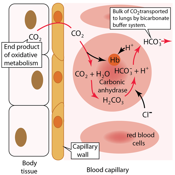
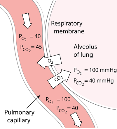
Oxygen in the lungs diffuses into the pulmonary capillaries and into red blood cells to bind to hemoglobin. This diffusion process of gas exchange depends upon the partial pressures and the solubility in the fluids and is described in more detail in Fick's Law, Graham's Law and Henry's Law

At the same time as the oxygen is preferentially diffusing into the blood, there is net diffusion of CO2 from the blood into the lungs to be exhaled.

Hemoglobin is an essential protein in the body, performing the major part of the oxygen transport in the blood. Hemoglobin contains iron which gives it a red color, and it contributes that color to the red blood cells and the blood itself.

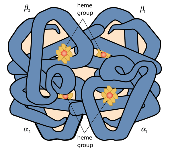
Hemoglobin consists of two types of components call heme and globin. Globin is a protein of 574 amino acids in four polypeptide chains. Each of those chains is associated with a heme group. Each heme group surrounds an atom of iron, and each iron atom can loosely bind an oxygen atom. Binding up to four oxygen molecules, the hemoglobin forms the compound called oxyhemoglobin. Myoglobin consists of one polypeptide chain similar to one of the four in the structure above, and can bind only one oxygen molecule. It is abundant in muscle cells and acts as a storage location for oxygen which the cell calls upon in times of low oxygen supply.

Any animal greater than a few millimeters in size must ensure a steady supply of oxygen to cells throughout its body and remove waste products such as carbon dioxide. In all vertebrates and even in some microscopic life the transport protein is hemoglobin. Myoglobin is also used by most animals in muscle tissue to provide an oxygen reserve for periods of high oxygen demand.

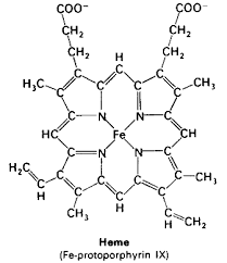
An iron atom in the state FeII forms an iron porphyrin which provides the vivid red color for blood and red blood cells. Plants use a magnesium porphyrin in chlorophyll which gives plants their green color.

This iron porphyrin binds to the proteins to provide the oxygen binding site.

A delicately balanced binding site for oxygen

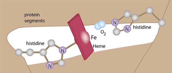
The details of the polypeptide environment of both myoglobin and hemoglobin bind the oxygen but protect the iron from oxidation. This exquisitely balanced environment allows the oxygen to bind and be released so that it can bind another oxygen. The framework for holding the heme involves two histidines which help stabilize the heme in a pocket of the globular structure of either myoglobin or hemoglobin.

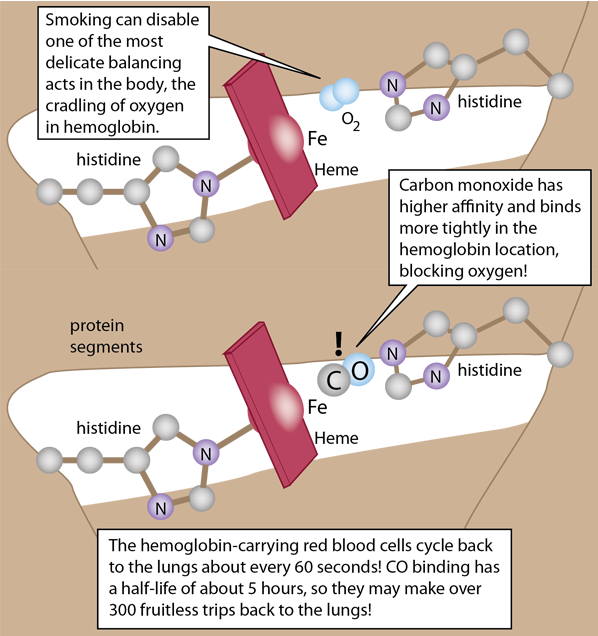
The heme pocket is ideal for oxygen, but carbon monoxide actually binds to both hemoglobin and myoglobin with much greater affinity and that binding is not readily reversible. This makes CO a potent poison in the body.

When provided with a supply of oxygen, myoglobin will bind oxygen rapidly even at very low partial pressures of oxygen. That makes it efficient as a storage location for oxygen, but this kind of affinity curve makes it a poor supplier of oxygen at typical partial pressure values in tissue. Hemoglobin's affinity starts low and rises gradually up to a supply partial pressure of about 20 mmHg, but then turns upward sharply. This change in its behavior (an allosteric effect) comes from the fact that the binding of an oxygen molecule to the first of the four hemes causes a configuration change in hemoglobin that makes it easier for it to bind oxygen to the other three hemes. This is called "cooperative binding" and it is of tremendous importance in the efficiency of oxygen transport in animals.

Myoglobin in tissue - especially in muscle tissue - accepts oxygen from hemoglobin and stores it. It can then deliver the oxygen to the mitochondria when their oxygen needs are sufficiently great.

The Bohr Effect

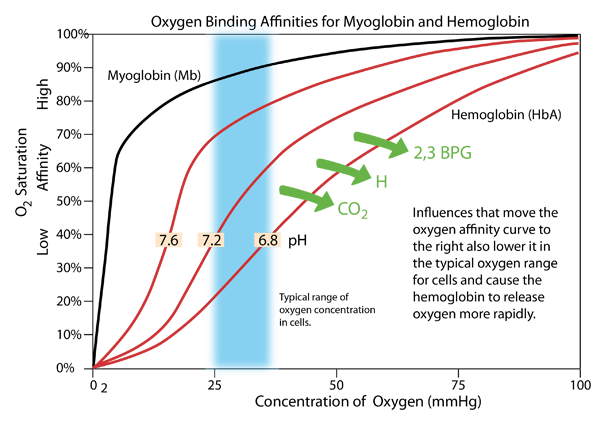
The pH of the blood is close to the neutral pH of 7.4, typically ranging from 7.35-7.45 and needing to be maintained in that range. But inside cells the pH can vary over a wider range, and the pH affects the oxygen supply process. If the pH becomes lower (more acidic), the oxygen affinity curve of hemoglobin moves to the right as shown above, and exhibits lower affinity in the range of oxygen concentration typical of the cellular interior. Active metabolism in the cell produces CO2 and H, which act to lower the pH. This causes oxygen to be released more rapidly in an effect known as the Bohr Effect, named after Christian Bohr who discovered this phenomenon.

An additional effect is the binding of the compound 2,3 bisphosphoglycerate (2,3 BPG) which enhances the stability of the deoxyhemoglobin and also has the affect of moving the affinity curve to the right, enhancing oxygen release.

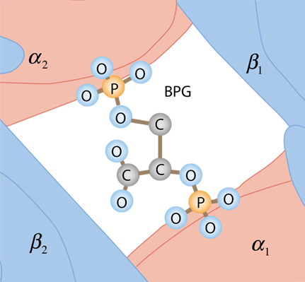
The BPG is bound in the center space between the four globulins of deoxyhemoglobin by the surface charges on the proteins. It does not fit in this space for oxyhemoglobin, so it does not bind.

Index

Biochemical concepts

Chemistry concepts

Reference
Shipman, Wilson and Todd
Ch 15

Matthews, van Holde, Ahern
Ch 7

Ahern
Ch 6
 
HyperPhysics*****Chemistry R Nave
Go Back


Fetal Hemoglobin

A remarkable aspect of the hemoglobin oxygen delivery system is the difference between fetal hemoglobin and normal adult hemoglobin. These differences increase the oxygen affinity of fetal hemoglobin and allow the fetus to extract oxygen from the normal hemoglobin of the mother in the placental circulation. One of the reasons for the higher oxygen affinity of the fetal hemoglobin is that it does not bind 2,3 BPG as strongly as the mother's adult hemoglobin.

The structure of fetal hemoglobin differs from adult hemoglobin which has an α2β2 structure by replacing one pair with γ chains to produce an α2γ2 structure. This fetal hemoglobin has a much lower affinity for BPG than the adult version and will therefore have a higher oxygen affinity. This difference which allows the fetus to gain oxygen from the mother's blood is accomplished by replacing a pair of histidines (His 143 according to Matthews, et al.) in the adult hemoglobin with a pair of serines in location 143 (replacing 2 out of 574 amino acids). Wiki suggest either an alanine or a glycine at position 136.

Fetal hemoglobin wiki
Index

Biochemical concepts

Chemistry concepts

Reference
Shipman, Wilson and Todd
Ch 15

Matthews, van Holde, Ahern
Ch 7

Ahern
Ch 6
 
HyperPhysics*****Chemistry R Nave
Go Back


Smoking and Hemoglobin

One of the major health risks of smoking has to do with the binding of carbon monoxide to hemoglobin, blocking the binding of oxygen and decreasing the oxygen supply to the tissues.

The article referenced below on carbon monoxide suggests the following data about carbon monoxide [this data has not been checked carefully for these comments]: "Carbon monoxide is much faster at binding with hemoglobin than oxygen (about 200 times faster)." "Carbon monoxide is quick to connect with red blood cells but is slow to exit the body, taking as much as a day to be exhaled through the lungs." "The normal level of COHb in the bloodstream from environmental exposure is less than 1%. ..A pack-a-day smoker can have 3% to 6% COHb in the blood. ... In a three pack-a-day smoker, COHb levels may reach 20%."

Medical News Today states that carbon monoxide clears from the body in 12 hours. An NIH paper suggests a half-life of 4.5 hours for carbon monoxide leaving the body after smoking.

Another factor that discriminates against oxygen uptake by the hemoglobin of smokers is the fact that they have higher levels of 2,3 BPG in their blood. Non-smokers have less BPG associated with their hemoglobin, and it tends to come off during the transit to the lungs and be broken down. By the time this hemoglobin reaches the lungs, very few hemoglobins have BPG so they have higher binding affinity for oxygen.

Red blood cell wiki
Carbon monoxide and smoking
Index

Biochemical concepts

Chemistry concepts

Reference
Shipman, Wilson and Todd
Ch 15

Matthews, van Holde, Ahern
Ch 7

Ahern
Ch 6
 
HyperPhysics*****Chemistry R Nave
Go Back


Red Blood Cells


Scanning electron micrograph, credit NIH, Noguchi, Rodgers & Schechter.

Red blood cells, formally called erythrocytes, are abundant in the body and carry vast numbers of hemoglobin molecules to supply oxygen for body processes. Each red blood cell contains approximately 270 million hemoglobin molecules. They are 6-8μm in diameter. These cells lack a cell nucleus and most of the organelles of normal body cells in order to accommodate maximum space for hemoglobin. The cell membranes are stable, but very deformable so that they can traverse the circulatory system including the capillary network. Adults produce about 2.4 million new erythrocytes each second. These cells develop in the bone marrow and circulate for 100-120 days in the body. They make a circuit of the circulatory system from the lungs and back in about 60 seconds. Approximately 84% of the cells in the human body are the 20-30 trillion red blood cells. Nearly half of the blood's volume (40% to 45%) is red blood cells. (Wiki)

The red blood cells contain the enzyme carbonic anhydrase to help convert CO2 to H2CO3 for transport of the metabolic product CO2 to the lungs for exhalation.

In response to low blood oxygen levels, the kidneys release the hormone erythropoietin, which travels in the blood to the bone marrow. There, it stimulates more rapid production of the red blood cells that contain hemoglobin to transport oxygen to the cells.

Red blood cell wiki
Index

Biochemical concepts

Chemistry concepts

Reference
Shipman, Wilson and Todd
Ch 15

Matthews, van Holde, Ahern
Ch 7

Ahern
Ch 6
 
HyperPhysics*****Chemistry R Nave
Go Back


Carbon Dioxide Transport to Lungs

The body's metabolic processes such as the reactions in the TCA cycle release carbon dioxide into the body fluids. This CO2 must be excreted from the body by transporting it to the lungs to be exhaled. The active metabolism in the cell produces CO2 and it reacts with water to form bicarbonate ions HCO-3 and hydrogen ions H+. As described with the Bohr effect above, this process lowers the pH of the cell and causes more oxygen to be released. In this way, the CO2 helps to provide the necessary oxygen for the cell's metabolic demands.

At some point, the excess CO2 must be removed to maintain a proper balance in the cell. Whereas about 98.5% of the oxygen supply to the cells is carried by hemoglobin compared to 1.5% dissolved in the blood, the transport of CO2 is profoundly different. Carbon dioxide is more soluble than oxygen as a gas in blood, and about 5-7% of it is dissolved in the blood to be carried to the lungs. About 10% of the CO2 binds to plasma proteins or enters the red blood cells to bind to hemoglobin for transport.

When carbon dioxide binds to hemoglobin it first forms bicarbonate ions HCO-3 and hydrogen ions H+ as described above and then binds to one of the amino groups of hemoglobin:

With this structure bound to hemoglobin, the molecule is called carbamohemoglobin, the carrier molecule to the lungs. The binding of this structure is reversible, and can freely disassociate from the hemoglobin when it reaches the lungs and quickly release CO2 for exhalation.

The majority of carbon dioxide molecules (85%) are carried to the lungs by the bicarbonate buffer system. Carbon dioxide diffuses into the red blood cells where the enzyme carbonic anhydrase (CA) quickly converts it to carbonic acid (H2CO3). Carbonic acid is an unstable intermediate molecule that immediately dissociates into bicarbonate ions. This is a steep favorable path from CO2 to HCO-3 down its concentration gradient so that CO2 can continue to enter the red blood cells. This also produces H+ ions, which if allowed to accumulate would produce dangerous changes in the tightly regulated pH of the blood. The role of the hemoglobin in the red blood cells here is to bind these H+ ions, limiting their effect on the pH. The newly synthesized bicarbonate ions are transported out of the red blood cells into the liquid content of the blood in exchange for a chloride ion Cl-. (This is called the chloride shift.) When the blood reaches the lungs, the bicarbonate is transported back inside the red blood cells in exchange for the Cl-. The H+ ions disassociate from the hemoglobin and bind to the bicarbonate, producing the intermediate carbonic acid. This carbonic acid is converted back to CO2 by the action of the enzyme carbonic anhydrase and diffuses into the alveoli of the lungs for exhalation.

The remarkable accomplishment of this chain of events is to transport large mounts of CO2 from the cell metabolism where they are released to the lungs where they are exhaled with very little change in the pH of the blood. Note that in the discussion of the Bohr effect there is discussion of pH changes from about 7.6 to 6.8 inside of cells, but the pH of the blood itself is limited in range from about 7.35 - 7.45 with serious to fatal consequences for excursions outside that range. This key enzyme, carbonic anhyrase, accomplishes what is called the bicarbonate buffer system which you make use of with every breath you take.

Gas transport in blood:opentextbc.ca
Carbonic anhydrase wiki
Index

Biochemical concepts

Chemistry concepts

Reference

Matthews, van Holde, Ahern
Ch 7

Ahern
Ch 6

News-medical
 
HyperPhysics*****Chemistry R Nave
Go Back