The Urinary System is One of the Excretory Systems
|
Index Reference Audesirk & Audesirk Ch 26, 30 Thibodeau & Patton Ch 28 | |||||||||||||||||
|
Go Back |
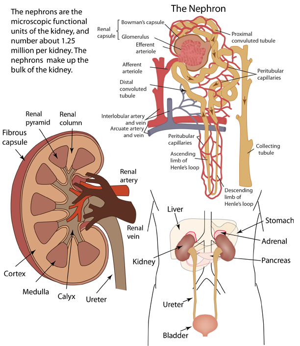
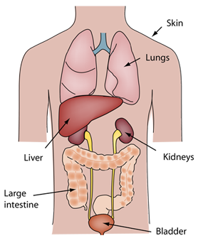
The Urinary SystemThese sketches of the elements of the urinary system are patterned after the treatment by Thibodeau & Patton. |
Index Reference Audesirk & Audesirk Ch 26, 30 Thibodeau & Patton Ch 28 | ||
|
Go Back |
The Urinary System and HomeostasisWalter B. Cannon is credited with introducing the term "homeostasis" in the 1920s to describe the constancy of the body's internal environment. Though "homeostasis" from the Greek implies that it "stays the same", the body is clearly a very active, ever-changing environment. The value of the term is to describe the dynamic equilibrium of many interacting systems in the body which result in a reasonably balanced functional organism. There are a number of physical and chemical parameters in the body that must be kept within a very narrow range for the cells to function properly. The kidneys and the urinary system are important parts of the homeostatic balance of the body. While we may think of the kidneys primarily as a "urine producer", Thibodeau & Patton suggest that a better image of the system would be that of "blood plasma balancer". Audesirk & Audesirk suggest a list of the ways that the urinary system helps maintain the homeostasis of the body:
The kidneys are the most important organs in the body for maintaining fluid-electrolyte and acid-base balance. "Each drop of blood in your body passes through a kidney about 350 times a day; thus the kidney is able to fine-tune the composition of the blood and thereby helps maintain homeostasis."(Audesirk & Audesirk) |
Index Reference Audesirk & Audesirk Ch 26, 30 Thibodeau & Patton Ch 28 | ||
|
Go Back |
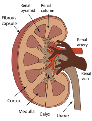
Elements of the Urinary SystemKidney Structure
Renal Pelvis: The larger collection reservoir formed by the joining of the calyx units. This reservoir narrows as it exits the kidney to become the ureter. Renal artery and Renal vein: These blood vessels are quite large and carry about 1200ml of blood per minute, or about one fifth of the total blood flow from the heart. Between the pyramids of the kidney's medulla, the renal artery branches to form interlobular arteries that extend outward toward the cortex and arch over the bases of the pyramids to form the arcuate arteries. From these arcuate arteries, interlobular arteries extend into the cortex. The Nephron
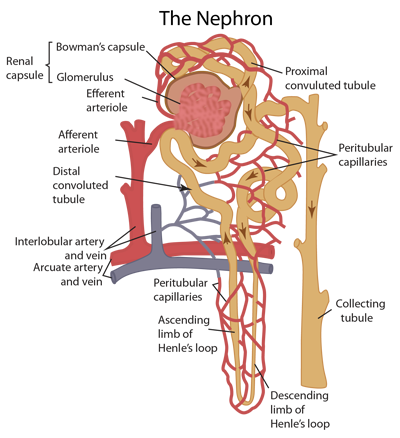
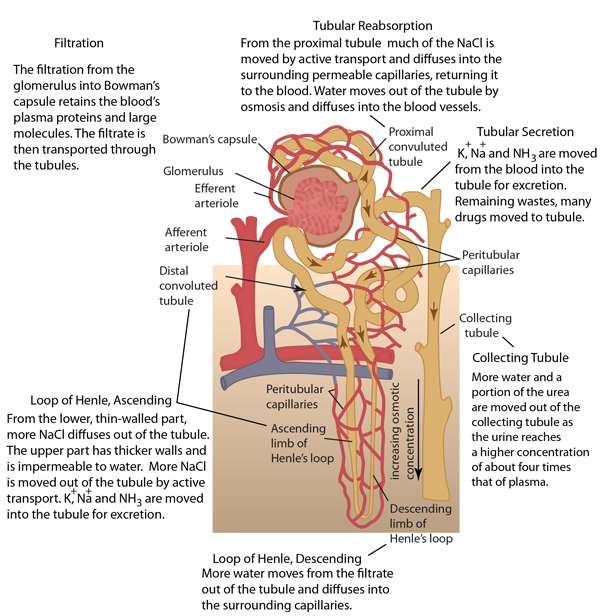
Afferent arteriole: From an interlobular artery, blood enters the afferent arteriole which carries the blood into the glomerulus for filtering. Efferent arteriole: After passing through the glomerulus, the remaining fluid with the filtrate removed is much more concentrated and passes into the smaller efferent arteriole and branches into multiple small, highly porous capillaries. These capillaries surround the tubule which has carried the filtrate from Bowman's capsule and are in intimate contact with it. Proximal convoluted tubule: the filtrate leaving Bowman's capsule into the tubule contains nutrients and water and a portion of those are reabsorbed into the blood flowing through the nearby capillaries. This reabsorption can include salts, glucose, and amino acids which are excreted from the tubule by active transport and then diffuse into the porous capillaries. At the same time, remaining waste products including urea become more concentrated in the tubule. Loop of Henle: just past the proximal tubule, a thinner tubule descends to a sharp turn and returns with an enlarged tubule. This structure extends into the medulla of the kidney for most nephrons. Along the loop of Henle there is more concentration of the urine by osmosis of water out of the tube and transfer of NaCl out by active transport. Distal convoluted tubule: Beyond the loop of Henle the tubule moves to a point adjacent to the afferent arteriole and the two of them form the juxtaglomerular apparatus. This structure is important in maintaining homeostasis of blood flow because it reflexively secretes renin when the blood pressure in the afferent arteriole drops. Renin triggers a mechanism that produces angiotensin, a substance that causes vasoconstriction and a resulting increase in blood pressure. Collecting tubule: From the distal tubule the urine enters the straight tubule that is joined by the distal tubules of several nephrons.
|
Index Reference Audesirk & Audesirk Ch 26, 30 Thibodeau & Patton Ch 28 | ||||||
|
Go Back |
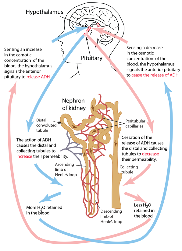
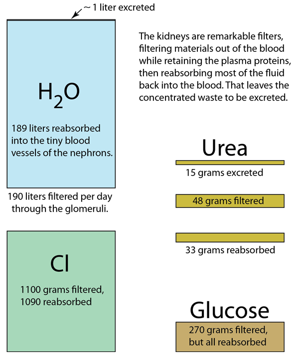
Regulating the Water Content of the BloodOne of the most important functions of the kidney is to regulate the water content of the blood. Audesirk & Audesirk comment that the kidneys filter about 125 ml (about half a cup) of water per minute, so that without reabsorption you would produce more than 180 liters (45 gallons) of urine daily! The reabsorption into the blood occurs by osmosis from the convoluted tubules and collecting tubules of the nephrons. The water can then diffuse into the permeable capillaries of the nephron which surround the tubules. The amount or reabsorption is controlled by a negative feedback mechanism that involves the amount of antidiuretic hormone (ADH) circulating in the blood. This control process involves the endocrine system and involves the hypothalamus which detects the osmotic concentration of the blood. Also involved are receptors in the heart that monitor blood volume. In response to increased osmotic concentration, the hypothalamus signals the pituitary gland to release ADH into the blood to increase the permeability of the tubules of the nephrons. |
Index Reference Audesirk & Audesirk Ch 26, 30 Thibodeau & Patton Ch 28 | ||
|
Go Back |
Fluid Exchange Processes in the NephronMultiple processes in the nephrons of the kidney accomplish the balancing of the constituents of the blood and interstitial fluid while concentrating the urine for excretion. In addition to the overall regulation of the water content of the blood, the complex process retains the plasma proteins and large molecules in the blood. It also regulates nutrients such as glucose, amino acids, vitamins, urea and a variety of ions including sodium, potassium, chloride and sulfate. In this context there are several descriptive terms: Filtration: The main part of the filtration of the blood occurs in the glomerulus of the nephron, with the resulting fluid called the filtrate collected in Bowman's capsule and then proceeding to the tubules. The filtrate subsequently refers to the content of the tubules and further structures leading to the final excretion of the urine. Tubular Reabsorption: The process by which cells of the proximal tubule remove water and nutrients from the filtrate within the tubule and pass them back into the blood. Tubular Secretion: the process by which wastes and excess substances that were not initially filtered out into Bowman's capsule are removed from the blood for excretion. The multiple exchange processes allow the kidney to maintain the needed constituents in the blood with appropriate balance and also to excrete urine that is about four times the molar concentration of the blood plasma. A significant contributor is the fraction of nephrons which have long loops of Henle that extend down into the medulla of the kidney. The fluid exchange processes of the lower parts of the loop lead to an increasing concentration. Audesirk & Audesirk comment on the example of the desert kangaroo rat which has only very long loops of Henle and is able to concentrate its urine to 14 times its plasma concentration! The tubular secretion process associated with the distal tubule is credited with helping remove a number of substances. Hydrogen ions are removed from the blood by active transport there, and the kidney's regulation of the pH of the blood is associated with control of the relative concentration of hydrogen and bicarbonate ions. The kidney removes some potential harmful substances such as some drugs, pesticides, food additives, and nicotine from cigarette smoke. |
Index Reference Audesirk & Audesirk Ch 26, 30 Thibodeau & Patton Ch 28 | ||
|
Go Back |
Filtration and Reabsorption by the KidneyThe kidneys handle many substances in their role of contributing the fluid balance and homeostasis of the body. This data is from Thibodeau & Patton. Almost all of the useful cellular constituents are reabsorbed into the blood. The urea cycle in the liver converts the toxic NH3 into urea, a non-toxic form which is then transported to the kidneys. One might expect that the urea would all be excreted, but in fact only about a third of it is excreted. The reabsorption of urea provides a higher concentration of urea in the inner medulla of the kidney and is thought to contribute to being able to achieve the observed concentration of the urine in the collecting tubule. (Ask a Biologist) |
Index Reference Audesirk & Audesirk Ch 26, 30 Thibodeau & Patton Ch 28 | ||
|
Go Back |