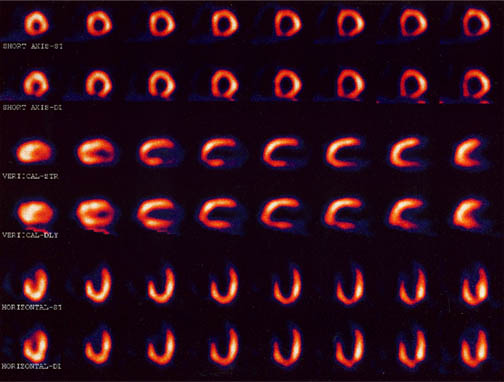
Myocardial Infusion Imaging
A widely used diagnostic procedure for heart health is the imaging of the blood infusion to the heart using a low dose of a radioactive tracer in the blood. A large radiation detector rotates about the chest of the patient, detecting gamma radiation from the tracer element which has been injected into the patient's vein. The image shows the perfusion of the blood into the heart muscle.
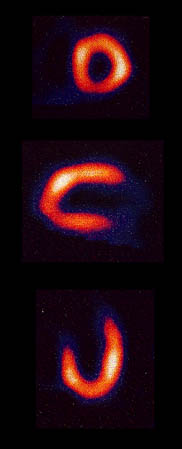 |
The detection of the gamma rays produces images of the blood flow in the heart muscle from various angles to provide an assessment of the infusion of blood. Early tests of this type used an isotope of thallium with a gamma ray of energy about 70 keV. This kind of image came to be known as a "thallium scan", but now an isotope of technetium is the isotope of choice for the scans, having a shorter halflife and producing a gamma of energy about 140 keV. |
The large detector uses an ionization detection in a manner similar to a Geiger counter and collects a series of exposures as it sequences around the body trunk. A collection of images is illustrated below, taken before and after exercise on a treadmill for comparison of the infusion of the blood under those two conditions.
Nuclear Medicine
| HyperPhysics***** Nuclear | R Nave |